 |
Oxidative Stress
Rev.081027 |
Anti-8-OHdG Monoclonal Antibody (clone N45.1)
Anti- 8-hydroxy-2'-deoxyguanosine monoclonal antibody |
|
| About 8-hydroxy-2'-deoxyguanosine (8-OHdG): |
 |
Reactive oxygen species (ROS) and oxidative stress may be crucial for development of various diseases such
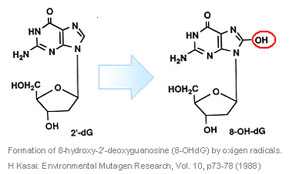
as diabetes, cancer, and may play an important role in the aging process. 8-hydroxy-2'-deoxyguanosine (8-OHdG) is a product of
oxidatively damaged DNA formed by hydroxy radical, singlet oxygen and direct photodynamic action. 8-OHdG can be detected in
tissue, serum, urine and other biomaterials. Anti 8-OHdG monoclonal antibody (clone N45.1) is highly specific for DNA damage,
not cross react with RNA oxidation products such as 8-hydroxy-guanine and 8-hydroxy-guanosine.
Suitable for immunohistochemistry and ELISA.
|
 |
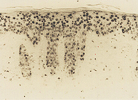
Epidermis from hairless mouse by chronic UVB irradiation after 4 weeks of treatment
stained with N45.1.
(Y.Hattori, et.al.: J Invest Dermatol 107, p733-737, 1996) |
|
| Specifications |
| Clone #: |
N45.1 |
| Antigen: |
8-OHdG-conjugated Keyhole Limpet Hemocyanin |
| Subclass: |
Mouse IgG1(kappa)
Prepared as ascite, and ammnonium sulphate purified. |
| Form: |
Lyophilized Powder |
| Package: |
20 or 100 µg of IgG / vial (MOG-020P and MOG-100P respectively). |
| Specificity: |
19 analogues of 8-OHdG {guanosine (G), 7-methyl-G, 6-SH-G, 8-bromo-G, dA, dC, dT, dI, dU, dG, O6-methyl-dG, 8-OHdA,
guanine (Gua), O6-methyl-Gua, 8-OHGua, uric acid, Urea, creatine, creatinine} demonstrate no cross-reactivity.
Only 8-sulfhydryl-G and 8-OHG demonstrate minimal cross-reactivity (less than 1%). |
| Use: |
Immunohistochemistry and ELISA |
|
| Product name |
Code |
Content |
| Anti 8-OHdG monoclonal antibody |
MOG-020P |
20 µg of IgG |
| MOG-100P |
100 µg of IgG |
|
|
| FOR RESEARCH USE ONLY. Not for diagnostic, medical or other use. |
| 1) |
S.Toyokuni, T.Tanaka, Y.Hattori, Y,Nishiyama, A.Yoshida, K.Uchida, H.Hiai, H.Ochi, and T.Osawa:
Quantitative immunohistochemical determination of 8-hydroxy-2'deoxyguanosine by a monoclonal antibody N45.1:
Its application to ferric nitrilotriacetate-induced renal carcinogenesis model.
Lab. Invest. 76(3), p365-374 (1997)
[ Specifity of the antibody clone N45.1.] |
| 2) |
Y.Hattori, C.Nishigori, T.Tanaka, K.Uchida, O.Nikaido, T.Osawa, H.Hiai, S.Imamura, and S.Toyokuni:
8-Hydroxy-2'deoxyguanosine is increased in epidermal cells of hairless mice after chronic ultraviolet B exposure.
J. Invest. Dermatol. 107, p733-737 (1996)
[ Immunohistochemistry of UV-B irradiated mouse skin. ] |
| 3) |
T.Tanaka, Y.Nishiyama, K.Okada, K.Hirota, M.Matsui, J.Yodoi, H.Hiai, and S.Toyokuni:
Induction and nuclear translocation of thioredoxin by oxidative damage in the mouse kidney: independence of tubular necrosis and
sulfhydryl depletion. Lab. Invest. 77(2), p145-155 (1997)
[ Immunohistochemistry of ferric nitrotriacetate-treated mouse kidney. ] |
| 4) |
Y.Ihara, S.Toyokuni, K.Uchida, H.Odaka, T.Tanaka, H.Ikeda, H.Hiai, Y.Seino, and Y.Yamada:
Hyperglycemia causes oxidative stress in pancreatic -cells of GK rats, a model of type 2 diabetes.
Diabetes 48, p927-932 (1999)
[ Immunohistochemistry of pancreatic islets from GK rat, a diabetes model. ] |
| 5) |
T. Ichiseki, A. Kaneuji, S. Katsuda1, Y. Ueda,T. Sugimori and T. Matsumoto:
DNA oxidation injury in bone early after steroid administration is involved in the pathogenesis of steroid-induced osteonecrosis.
Rheumatology 44,p456-460(2005)
[ Immunohistochemistry of rabbit tissue. ] |
| 6) |
Lee YA, Cho EJ, Yokozawa T:
Protective effect of persimmon (Diospyros kaki) peel proanthocyanidin against oxidative damage under H2O2
-induced cellular senescence. Biol Pharm Bull 31(6)p1265-1269(2008)
[ Application to human cultured cells. ] |
|
| Immunohistochemical procedure |
| Fixation: |
For formalin-fixed tissue and cultured cells, antigen retrieval process may be useful.
Tissue samples are fixed by Bouin¡Çs Solution, antigen retrieval process will not be needed. |
| Blocking: |
Apply normal rabbit serum (Diluted to 1:75) (DAKO corporation, Kyoto, Japan) to the specimens
for the inhibition of non-specific binding of secondary antibody.
If sample tissues are from mouse, blocking of internal mouse IgG will be needed. |
| Primary antibody: |
Apply N45.1 (1-10microgram/ml) to the specimens, incubate overnight at 4 degree C. |
| Secondary antibody: |
Apply biotin-labeled rabbit anti-mouse IgG serum (Dako; Diluted to 1:300) for
40min at room temperature. |
| ABC method: |
Apply avidin-biotin-alkaline phosphatase complex (Vector Lab., Burlingame, CA; Diluted to 1:100)
for 40min at room temperature. |
| Staining: |
Use a kit of black substrate for alkaline phosphatase (BCIP/NBT, Vector). |
| |
Antigen retrieval:
Put sample slides into 500£íL glass beaker with 10mM citrate buffer pH6.0 and heat.
Keep boiling for 5 minutes.
Cool down slowly in 1 hour.
|
|
 |
| FOR RESEARCH USE ONLY. Not for diagnostic, medical or other use. |
|


